Orthopedic specialists rely on advanced imaging technologies to accurately diagnose bone and joint problems. These diagnostic tools provide detailed views of the musculoskeletal system. Understanding how these imaging methods work helps patients prepare for their appointments and comprehend their treatment plans.
Understanding Bone Structure Analysis
X-rays represent the most commonly used imaging technique by orthopedic specialists. These images provide clear views of bone structure, making them ideal for detecting fractures, bone deformities, and joint spacing issues. Orthopedic specialists can quickly identify broken bones, dislocations, and signs of arthritis through X-ray examination.
The X-ray process involves positioning the patient so that the affected area lies between an X-ray machine and a detector plate. The machine sends electromagnetic radiation through the body, and different tissues absorb varying amounts of this radiation. Bones appear white on X-ray images because they absorb more radiation than surrounding soft tissues.
Exploring Soft Tissue Evaluation
MRI (Magnetic Resonance Imaging) and CT (Computed Tomography) scans provide more detailed information about both bone and soft tissue structures. MRI uses powerful magnetic fields and radio waves to create detailed images of muscles, ligaments, tendons, and cartilage. This technology excels at showing soft tissue injuries, such as torn ligaments or damaged cartilage that X-rays cannot detect.
CT scans combine X-ray technology with computer processing to produce cross-sectional images of the body. These scans offer superior detail of bone structure compared to traditional X-rays and can reveal complex fractures, bone tumors, and subtle bone abnormalities. Orthopedic specialists frequently use CT scans when planning surgical procedures that require precise anatomical knowledge.
Ultrasound imaging provides real-time views of soft tissues. It can detect fluid accumulation, muscle tears, and tendon problems. This technique offers the advantage of dynamic imaging, allowing specialists to observe how structures move during examination.
Creating Treatment Plans
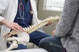
Orthopedic specialists analyze imaging results by examining bone density, joint alignment, soft tissue integrity, and overall structural relationships. They look for signs that indicate specific conditions or injuries. The interpretation process involves comparing the affected area to normal anatomy and identifying any deviations from expected patterns. Specialists may use measurements and angles from imaging studies to quantify the severity of conditions such as scoliosis or joint deformity.
Imaging results directly influence treatment decisions. Minor fractures visible on X-rays might require only immobilization and rest, while complex injuries shown on MRI or CT scans may necessitate surgical intervention. The detailed information provided by imaging studies allows specialists to develop targeted treatment plans that address each patient’s condition.
Find Orthopedic Specialists
Imaging technology plays a fundamental role in modern orthopedic diagnosis, providing specialists with the detailed information needed to identify bone and joint problems accurately. X-rays offer quick assessment of bone structure, while advanced techniques like MRI and CT scans reveal soft tissue damage and complex anatomical details. Understanding these diagnostic tools helps patients appreciate the thoroughness of their evaluation and the precision involved in developing their treatment plans. Discuss any questions about recommended imaging studies with your orthopedic specialist to fully understand your diagnosis and treatment options.
- Obligation Linéaire: Complete Guide to Meaning, Use, Finance & Law
- Tarnplanen: The Complete 2025 Guide to Smart Planning for Success
- Ryan Trahan Net Worth: Full Breakdown of His Income, Career & Success
- Serlig: Complete Guide to Meaning, Benefits, Uses, Lifestyle & Modern Wellness Approach
- Escapamento RD: Complete Guide, Performance, Types, Installation & Maintenance